Unlabeled Printable Blank Skull Diagram
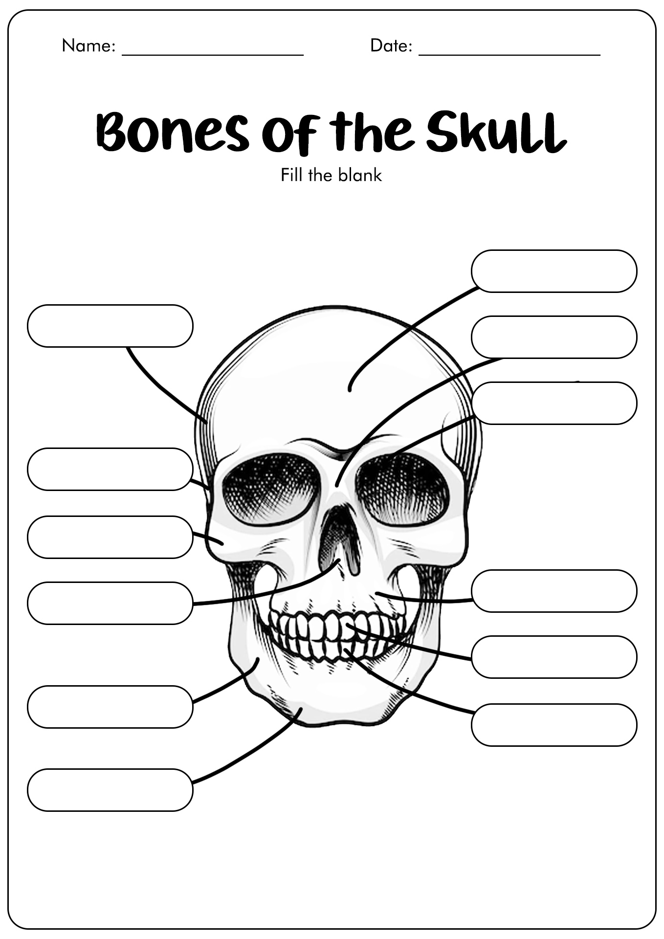
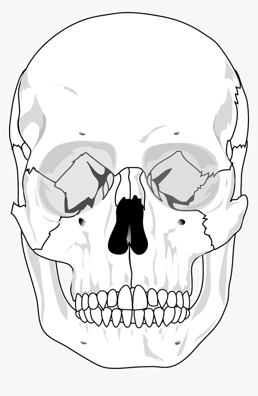
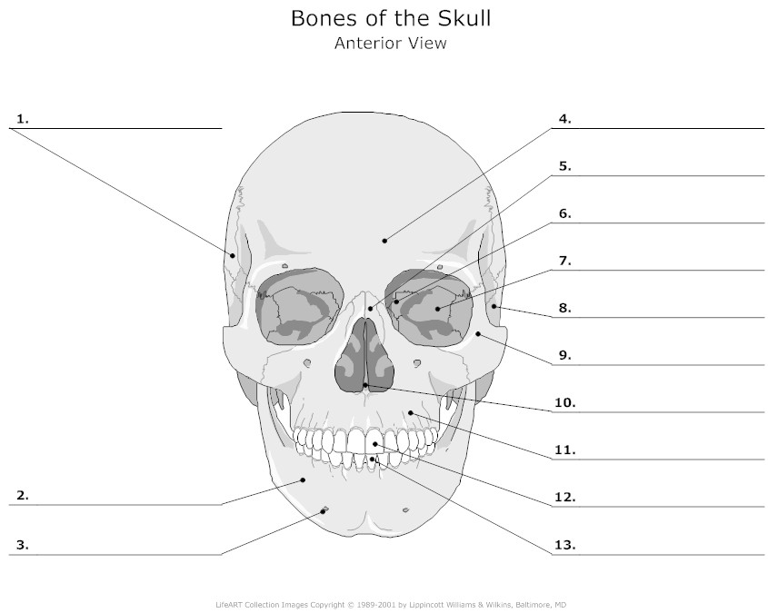
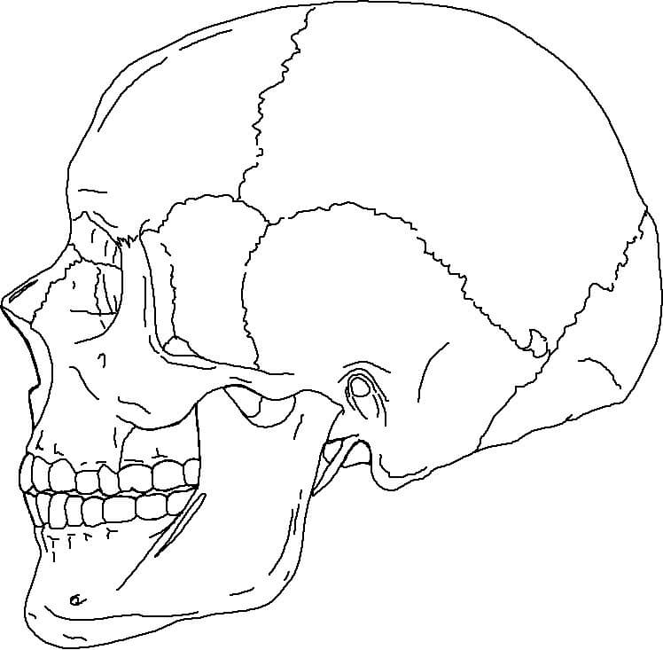
Unlabeled Printable Blank Skull Diagram - Define the paranasal sinuses and identify the location of each Define the paranasal sinuses and identify the location of each. This simple worksheet shows a skeleton with bones unlabeled. Color the bones of the hand. It even includes some of the carpal bones as well as the main bones of the body. Label and color the long bone. Locate and define the boundaries of the anterior, middle, and posterior cranial fossae, the temporal fossa, and infratemporal fossa. Pictures of skulls that are labeled for reference. Students fill in the boxes with the names of the bones. The axial and appendicular skeleton. How to use our free worksheet of the human skull for younger students: The parts of the skull have been labeled. Skeleton label using google slides. It allows the spinal cord to pass inferiorly out of the cranial vault, and also the vertebral arteries to enter the skull and provide the posterior input to the circle of willis. Mandible •. Here's how you can quickly and effectively refresh. Images adapted from images in the public domain. Web in this worksheet, we are going to review some of the major bones that protect and surround your brain. Web this diagram comes in six versions, all combined into one pdf: Web easy to download and print, use this activity at home to. Locate the major suture lines of the skull and name the articulating bones that form them; How many can you remember? Your challenge is to write the correct name for each part. Web the skeletal system is made up of more than 200 bones and has two main parts: Label and color the long bone. Locate the major suture lines of the skull and name the articulating bones that form them; Web foramen magnum (inferior view) just posterior to the middle of the skull is the foramen magnum.this is latin for large hole. You can also download the labeled version and use this to make some notes. Web this diagram comes in six versions, all. The skull consists of the rounded brain case that houses the brain and the facial bones that form the upper and lower jaws, nose, orbits, and other facial structures. You can also download the labeled version and use this to make some notes. Web the skeletal system is made up of more than 200 bones and has two main parts:. If you want to have more fun learning about bones, try our bone lab. Once you’ve done that, it’s time to learn anatomy with our skull labeling quiz. It's not always easy remembering the parts of the brain. Images adapted from images in the public domain. Color the bones of the foot. Here's how you can quickly and effectively refresh. Web print a few of these to quiz yourself. The skull consists of the rounded brain case that houses the brain and the facial bones that form the upper and lower jaws, nose, orbits, and other facial structures. This simple worksheet shows a skeleton with bones unlabeled. Images adapted from images in. Web print a few of these to quiz yourself. Print out our free skull worksheet that is already labeled. The anterior and posterior spinal arteries also. Great for artists and students studying human anatomy. Locate the major suture lines of the skull and name the bones associated with each. Color the bones of the foot. It's not always easy remembering the parts of the brain. Includes labeled human skeleton chart. You can also download the labeled version and use this to make some notes. The parts of the skull have been labeled. The mandible (lower jaw) is the largest strongest bone of the face and contains the. Includes labeled human skeleton chart. Web bones of the face. The skull consists of the rounded brain case that houses the brain and the facial bones that form the upper and lower jaws, nose, orbits, and other facial structures. Web the skeletal system is made. The axial and appendicular skeleton. How many can you remember? The mandible (lower jaw) is the largest strongest bone of the face and contains the. This simple worksheet shows a skeleton with bones unlabeled. How many skull bones are there, their structure, marking, & labeled pictures Locate the major suture lines of the skull and name the bones associated with each. Skeleton label using google slides. Web foramen magnum (inferior view) just posterior to the middle of the skull is the foramen magnum.this is latin for large hole. Foot (labeled) / charts and diagrams. Once you’ve done that, it’s time to learn anatomy with our skull labeling quiz. If you want to have more fun learning about bones, try our bone lab. Web our printable worksheet is free. Define the paranasal sinuses and identify the location of each It's not always easy remembering the parts of the brain. We’ve created a blank skull diagram free for you to download as a pdf below. Great for artists and students studying human anatomy.
Skull Unlabeled Anatomy and physiology, Skull, Anatomy
15 Best Images of Skull Labeling Worksheets Skull Bones Unlabeled
![[DIAGRAM] Lateral Of Skull Blank Diagram](http://www.ebmconsult.com/content/images/Anatomy/Skull_Anatomy_AP_view.png)
[DIAGRAM] Lateral Of Skull Blank Diagram
Skull Free Stock Photo Human Skull Diagram Blank, HD Png Download
4 Best Images of Diagram Of The Skull Printable Skull Bones Unlabeled

unlabeled skull Anatomy bones, Skull anatomy, Anatomy

Human Skull Diagram Blank , Free Transparent Clipart ClipartKey
31 Blank Skull Diagram To Label Labels Design Ideas 2020

Skull Bones Unlabeled Anatomy and physiology, Medical school

Printable Blank Skull Diagram
Locate And Define The Boundaries Of The Anterior, Middle, And Posterior Cranial Fossae, The Temporal Fossa, And Infratemporal Fossa.
They Will Practise Using Terms Such As:
You May Print The Skull Worksheet With Labels, Without Labels, Or Simply As A ‘Coloring Sheet’ Or Line Drawing Of The Human Skull.
It Even Includes Some Of The Carpal Bones As Well As The Main Bones Of The Body.
Related Post: